Lab 6: Coliform Determination by the Membrane Filter Technique
Lab 6: Lab Coliform Determination by the Membrane Filter Technique
Introduction
The purpose of this lab will ultimately be to familiarize ourselves with counting coliform microorganism colonies as well as another plating method called the membrane filter technique.
The total viable count is based upon the ability of a microorganism placed in a nutrient environment to grow. While individual microorganisms cannot be seen with the naked eye, they can grow in colonies which are visible to any plain Jane. Counting the colonies gives an indication of the number of viable colony forming units (CFU) present in the original sample. However, the appropriate growth medium must be chosen first to get to the counting phase. This part is crucial because the accuracy will be best if the chosen medium is as similar as possible to the sample environment.
The membrane filter technique uses the semi permeable membrane and a vacuum to trap the microorganisms and allow the rest of the solution to pass through the membrane pores. The membrane and its trapped microorganisms are then placed in a special plate with a pad saturated with the intended growth media
Two media will be used in this lab: M-Endo and M-FC. M-FC is a light pink growth media. It is a selective and differential media in that it only grows fecal coliform and the fecal coliform will grow blue (in the case that something else does grow). M-Endo is a dark pink media that will grow total coliform as 'gold' and is comprised of 1.0 % peptone, 0.25 % dipotassium hydrogen phosphate (K2HPO4), 1.0 % lactose, 0.33 % anhydrous sodium sulfite (Na2SO3), 0.03 % fuchsine and 1.25 % agar.
Methods and Materials
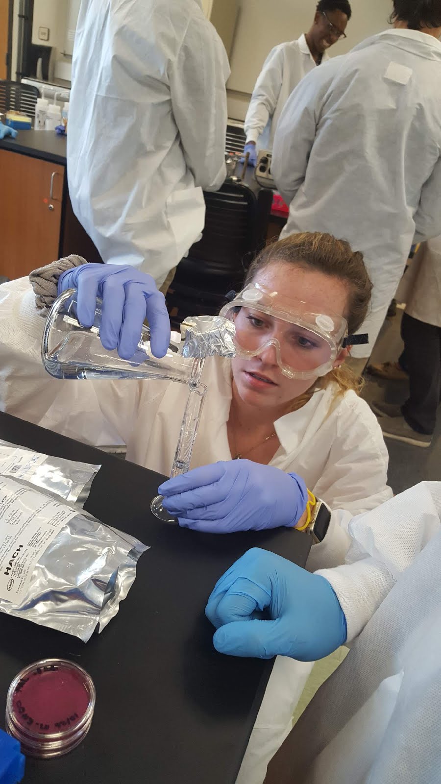
A sample of canal water was first chosen as the sample. A series of dilutions was first prepared, 10^-1 and 10^-2 in hopes of landing in between the range of acceptable 20-80 colony range. We ended up preparing 6 plates total, 2 plates of the original sample, 2 plates of 10^-1 and 2 plates of 10^-2. We plated on M-Endo for total coliform. Once the dilutions were made, the plates were prepared by adding the premeasured broth solution to the plates that had sterile pads. Then, the filter was placed onto the vacuum apparatus with the grid side facing up using a sterile pair of tweezers. 10 mL of the 10^-2 dilution was then added to the filter funnel and the suction was turned on. At the completion of the filtration, the cup was removed and the filter was carefully 'rolled' onto the media pad with the grid side facing up to avoid air entrapment. The funnel was then rinsed with DI water. This was repeated for the remaining 5 plates. Each plate was then inverted and allowed to incubate as follows: M-Endo at 35*C for 22-24 hours and M-FC at 44.5*C for 22-26 hours. After incubation, colonies were then counted and tabulated.
Results
Table 1. Dilutions, colony counts, original concentration and average concentration for M-Endo.

Sample Calculation: M-Endo, Plate 1, original sample.

All in all, probably shouldn't kill all the colonies. And we also shouldn't try to complete a lab in 15 minutes...this was not one of our more successful endeavors.
References
Nease, Steve. Fecal Coliform Bacteria at Play [Cartoons (Commentary)]. Accessed online 30 October 2017 at <http://images.oakville.halinet.on.ca/2822854/data?dis=ex>.
Thomson, Ashley. "Lab #6: Coliform Determination by the Membrane Filter Technique."
 |
| (Photo: Steve Nease) |
The purpose of this lab will ultimately be to familiarize ourselves with counting coliform microorganism colonies as well as another plating method called the membrane filter technique.
The total viable count is based upon the ability of a microorganism placed in a nutrient environment to grow. While individual microorganisms cannot be seen with the naked eye, they can grow in colonies which are visible to any plain Jane. Counting the colonies gives an indication of the number of viable colony forming units (CFU) present in the original sample. However, the appropriate growth medium must be chosen first to get to the counting phase. This part is crucial because the accuracy will be best if the chosen medium is as similar as possible to the sample environment.
The membrane filter technique uses the semi permeable membrane and a vacuum to trap the microorganisms and allow the rest of the solution to pass through the membrane pores. The membrane and its trapped microorganisms are then placed in a special plate with a pad saturated with the intended growth media
Two media will be used in this lab: M-Endo and M-FC. M-FC is a light pink growth media. It is a selective and differential media in that it only grows fecal coliform and the fecal coliform will grow blue (in the case that something else does grow). M-Endo is a dark pink media that will grow total coliform as 'gold' and is comprised of 1.0 % peptone, 0.25 % dipotassium hydrogen phosphate (K2HPO4), 1.0 % lactose, 0.33 % anhydrous sodium sulfite (Na2SO3), 0.03 % fuchsine and 1.25 % agar.
 |
| (Photo: Amy He) |
 |
| (Photo: Amy He) |
Methods and Materials
A sample of canal water was first chosen as the sample. A series of dilutions was first prepared, 10^-1 and 10^-2 in hopes of landing in between the range of acceptable 20-80 colony range. We ended up preparing 6 plates total, 2 plates of the original sample, 2 plates of 10^-1 and 2 plates of 10^-2. We plated on M-Endo for total coliform. Once the dilutions were made, the plates were prepared by adding the premeasured broth solution to the plates that had sterile pads. Then, the filter was placed onto the vacuum apparatus with the grid side facing up using a sterile pair of tweezers. 10 mL of the 10^-2 dilution was then added to the filter funnel and the suction was turned on. At the completion of the filtration, the cup was removed and the filter was carefully 'rolled' onto the media pad with the grid side facing up to avoid air entrapment. The funnel was then rinsed with DI water. This was repeated for the remaining 5 plates. Each plate was then inverted and allowed to incubate as follows: M-Endo at 35*C for 22-24 hours and M-FC at 44.5*C for 22-26 hours. After incubation, colonies were then counted and tabulated.
 |
| (Photo: Amy He) |
Results
Table 1. Dilutions, colony counts, original concentration and average concentration for M-Endo.

Sample Calculation: M-Endo, Plate 1, original sample.

M-Endo Plates
 |
| (Photo: Lauren Lukasik) |
Discussion
First and foremost, it is important to acknowledge that we incubated all of the plates for was too long. While they were supposed to be incubated for around a day, ours were incubated from Thursday-Tuesday. Oops! So basically we killed everything. Our M-Endo plates were supposed to reflect our coliform colonies as gold specks but they were all black. (RIP) The M-FC plates didn't have anything grown on them at all. So for future experiments, we probably shouldn't dehydrate the bacteria.
Based upon our results, only the first plate of the 10^-1 dilution is acceptable in terms of the 20-80 colony count range. However, our plates are all out of whack. Due to dilution error or cross contamination or just frying the bacteria, our counts for the 10^-1 dilution are drastically different. The first plate is 25 colonies and the second plate is 100 colonies which is a tad of a difference. The 25 colony plate would be in-line with the rest of the data but we goofed up on the second plate. Since that error is present, it throws off our average original concentration calculations and makes no sense of them. Oops.
If the fecal coliform plates reflected any colonies at all, they would have probably all been unusable because the colonies counts would have been less than the acceptable 20-80. Because the M-FC would only grow fecal coliform, this acts almost as if it were another dilution, meaning even though the original sample was not diluted, it would plate as if it were a 10^-1 dilution.All in all, probably shouldn't kill all the colonies. And we also shouldn't try to complete a lab in 15 minutes...this was not one of our more successful endeavors.
Nease, Steve. Fecal Coliform Bacteria at Play [Cartoons (Commentary)]. Accessed online 30 October 2017 at <http://images.oakville.halinet.on.ca/2822854/data?dis=ex>.
Thomson, Ashley. "Lab #6: Coliform Determination by the Membrane Filter Technique."


Comments
Post a Comment